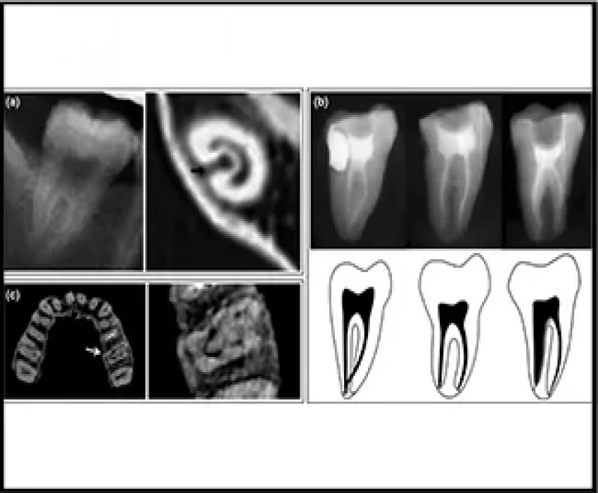
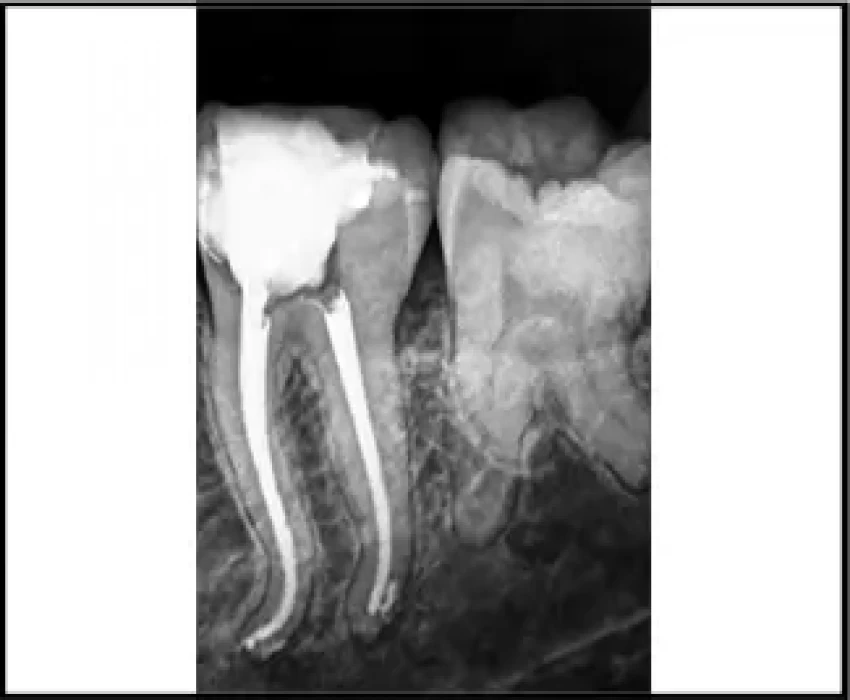
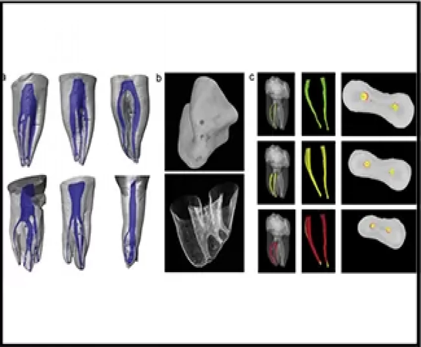
The pulp space is complex; root canals may divide and rejoin, and possess forms that are considerably more involved than commonly implied. Many roots have additional canals and a variety of canal configurations. Teeth with atypical canal configuration complicate the process of identifying and accessing root canals in the endodontic treatment.
The issue of prime importance with the treatment of atypical anatomies is undetected canals or incomplete debridement due to seemingly inaccessible root canals.
Whereas the skills and knowledge of the endodontist are paramount for successful root canal treatment, radiographic analysis is important for determining canal morphology. Cone-beam computed tomography (CBCT) and micro-CT can accurately determine root canal morphology. CBCT has the ability to overcome the limitations of conventional radiography such as three-dimensional evaluation of the complex canal anatomy during endodontic treatment. However, CBCT and micro-CT are not always feasible.
Combined X-ray analyses, however, such as performing the buccolingual view for identification of canal bifurcation and canal continuity, may increase the accuracy of identifying complex root canal morphology.
Periapical radiographs are still the most widely used method for determining root canal morphology before endodontic treatment. Accurate detection of complex canal morphology on X-ray is necessary to avoid missing root canals during treatment.
Following the SLOB rule in radiography, changing the horizontal angulation of the X-ray beam cone can, to a large extent, help in identifying and understanding the root canal anatomy. It makes it easier to place the newly found canal, lingual or buccal to the previously identified landmarks.
Another tact to understand root canal anatomies on radiographs can be:
- large canal becoming less obvious may logically determine there is a bifurcation;
- large canal becoming thinner and deviating towards one side, there may be one small and
- one large canal or furcated roots, and may logically determine that there is a bifurcation;
- medium root canal, gradual tapering, cannot logically determine that there are two canals but the proximal view may display a second root canal;
- buccolingual view shows a direct bifurcation.
Combined X-ray analyses, however, such as performing the buccolingual view for identification of canal bifurcation and canal continuity, may increase the accuracy of identifying complex root canal morphology.
Along with visualizing anatomy on the radiograph, clinical accessibility and visibility are much needed for complete debridement. Complete de-roofing of the pulp chamber along with the use of sodium hypochlorite for locating canal orifices are the most basic and simplest step to keep in mind to make our job easier and results, better. Magnification of the working field can enhance the visual fields multifold and improved visibility implies improved techniques. The Dental operating microscope is necessary for the detection of extra canals.
Various atypical anatomy calls for improvised techniques of debridement, preparation, and obturation to ensure a proper seal that is required for a successful endodontic treatment. An endodontic file should be used, which adapts to the natural morphology of the root canals and efficiently cleans it.
Along with improved clinical observation, a knowledge of common deviations from normal anatomy, their estimated frequency of occurrence can also help in better identification and treatment. Professionals should always consider morphological variations of the root canal system before the beginning of treatment.


