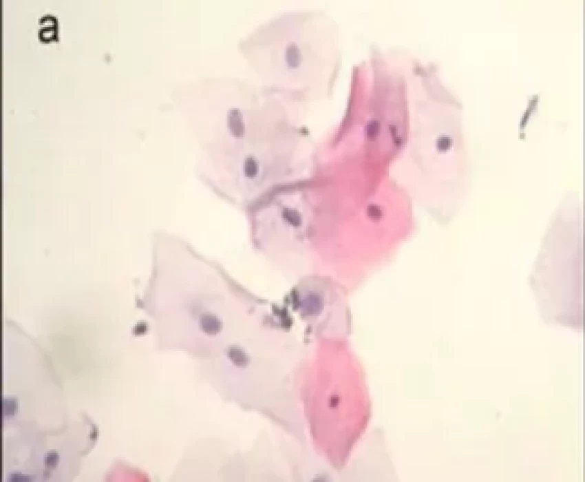
Smoking is currently the most inevitable cause of diseases and death worldwide and is one of the main risk factors for the development of oral cancer. Cigarette smoking in the development of oral malignancy has drawn increasingly interest. Therefore, it is mandatory to monitor the smoking patients carefully in view of the succession of alterations that smoking can cause. Exfoliative Cytology is one of the interesting tools by which one can screen the status of oral health in habit associated patients. Considering the possible influence of smoking on the occurrence of oral cancer and of precursor lesions, exfoliative cytology might be a useful tool for the detection and monitoring of initial alterations and for the establishment of adequate treatment in smokers. It is a complementary diagnostic method which presents several advantages such as rapid and easy execution, low cost, diagnostic safety, efficacy, and non-invasiveness, and can be repeated several times. Papanicolaou(PAP) staining is used as a routine method for the analysis of cytological aspects and permits the identification of basic inflammatory, dysplastic or malignant alterations. Ever since PAP described exfoliative cytology technique, which is a nonpainful, non-invasive procedure, it has become a valuable tool for cancer screening. The purpose of this study is to compare exfoliative cytology from oral mucosa of smokers and non-smokers, with an evaluation of proliferative activity by PAP (Papanicolaou) and AgNOR. Papanicolaou (PAP) staining is used as a routine method for the analysis of cytological aspects and permits the identification of basic inflammatory, dysplastic or malignant alterations. Ever since PAP described exfoliative cytology technique, which is a nonpainful, non-invasive procedure, it has become a valuable tool for cancer screening. Argyrophilic Nucleolar organizer regions (AgNORs) are located in the cell nucleoli during interphase. They are loops of DNA in which ribosomal RNA is encoded. Nucleolar organiser regions (NORs) are defined as nucleolar components containing a set of argyrophilic proteins, which are selectively stained by silver methods. After silver-staining, the NORs can be easily identified as black dots exclusively localized throughout the nucleolar area, and are called "AgNORs". The NORs' argyrophilic is due to a group of nucleolar proteins, which have a high affinity for silver (AgNOR proteins). the larger the number of NORs, the higher the replication rate of ribosomes and cells. This technique has therefore been used for the quantification of cell proliferation in different tissues and lesions.