
Orthodontic diagnosis has traditionally relied on clinical examination, radiographs, photographs, and dental stone casts. In recent years, three-dimensional (3D) digital study models-obtained through intraoral scanning or digitization of conventional casts-have significantly enhanced diagnostic accuracy and efficiency in orthodontics.
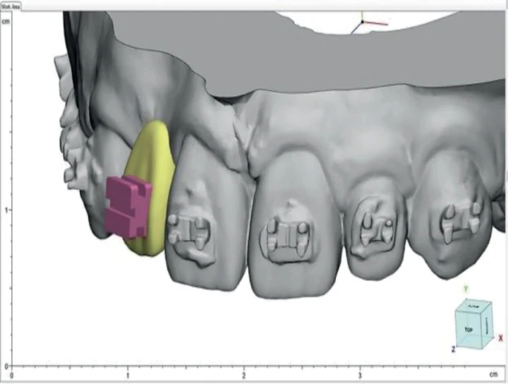
3D digital models provide a precise virtual representation of the dental arches, allowing clinicians to assess occlusion, tooth morphology, and arch form in all dimensions. Unlike physical casts, digital models can be magnified, and sectioned, offering improved visualization without the risk of damage or distortion.
A key advantage of digital models is the accuracy and reproducibility of orthodontic measurements. Common parameters such as mesiodistal tooth width, arch length discrepancy, inter-canine and inter-molar widths, overjet, overbite, and Bolton ratios can be measured reliably using digital software. Studies have shown these measurements to be comparable to those obtained from conventional stone models, with improved consistency due to ease of repeat measurements.
Digital models also facilitate integration with other diagnostic records such as cone beam computed tomography (CBCT) and facial scans, enabling a comprehensive three-dimensional evaluation of dental, skeletal, and soft tissue relationships. Furthermore, virtual treatment simulations assist in space analysis, appliance selection, and patient communication.
In summary, 3D digital models have become an integral component of modern orthodontic diagnosis by improving measurement accuracy, visualization, and interdisciplinary integration, thereby supporting more precise and efficient treatment planning.
No Any Replies to “How 3D Digital Models Are Changing Orthodontic Diagnosis”
Leave a Reply