
Assessment of Frontal Sinus, Maxillary Sinus Dimensions, and Nasal Septal Pattern in Gender Determination
Gender determination is a crucial aspect of forensic identification, particularly in cases involving fragmented or decomposed human remains. Craniofacial structures exhibit sexual dimorphism, and radiographic evaluation of paranasal sinuses and nasal septal patterns has gained importance as a reliable adjunct in forensic investigations. Among these, the frontal sinus, maxillary sinus, and nasal septum demonstrate individual variability and gender-related differences, making them valuable parameters for gender determination.
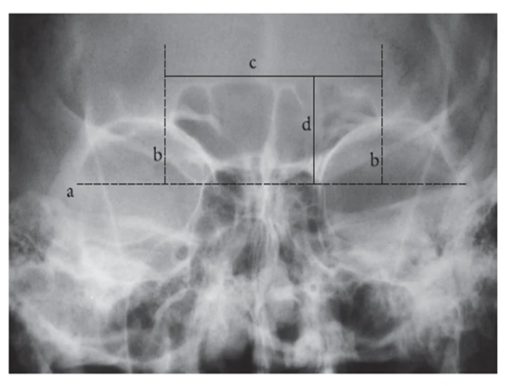
The frontal sinus is one of the most distinctive anatomical structures due to its unique morphology, even among monozygotic twins. Radiographic assessment of the frontal sinus includes evaluation of its height, width, area, symmetry, and overall contour. Studies have consistently shown that males tend to exhibit larger frontal sinus dimensions compared to females, attributed to greater craniofacial growth and hormonal influences. The frontal sinus is usually more pneumatized and asymmetrical in males, while females often show smaller, more symmetrical sinus patterns. This high degree of individuality enhances the forensic value of frontal sinus analysis.
Maxillary sinuses also exhibit sexual dimorphism and can be effectively assessed using panoramic radiographs, computed tomography (CT), or cone-beam computed tomography (CBCT). Measurements such as sinus height, width, depth, and volume are typically greater in males than females. The increased sinus volume in males is related to larger facial skeleton size and prolonged growth period. Maxillary sinus dimensions are particularly useful in forensic cases where the frontal sinus is absent, underdeveloped, or damaged. Three-dimensional imaging techniques allow precise evaluation of sinus morphology and improve accuracy in gender prediction.
The nasal septum, although less studied compared to paranasal sinuses, also shows gender-related morphological variations. The pattern of nasal septal deviation, thickness, and contour can provide supplementary information in gender determination. Males are more likely to exhibit pronounced septal deviations and thicker septal cartilage and bone, whereas females generally present with straighter or mildly deviated septa. While nasal septal patterns alone may not be definitive, their combined assessment with sinus morphology strengthens forensic conclusions.
The integration of frontal sinus, maxillary sinus dimensions, and nasal septal patterns enhances the accuracy of gender determination. Advanced imaging modalities such as CT and CBCT allow precise measurements and three-dimensional reconstruction, reducing observer variability and improving reproducibility. Statistical models and discriminant function analysis further aid in establishing gender with higher confidence.
However, it is important to recognize that sinus development is influenced by genetic, environmental, and ethnic factors. Pathological conditions, trauma, or surgical interventions can alter sinus morphology, potentially affecting accuracy. Therefore, radiographic findings should be interpreted in conjunction with other skeletal and dental parameters.
In conclusion, assessment of frontal sinus and maxillary sinus dimensions, along with nasal septal pattern analysis, serves as a valuable non-invasive tool in gender determination. When used in combination and supported by advanced imaging and statistical analysis, these parameters significantly contribute to forensic identification and strengthen the reliability of gender estimation.
No Any Replies to “Assessment of Frontal Sinus, Maxillary Sinus Dimensions, and Nasal Septal Pattern in Gender Determination:”
Leave a Reply