
CBCT-Based Evaluation of Bone Microarchitecture in Fibro-Osseous Lesions Using Image Analysis
Fibro-osseous lesions (FOLs) of the jaws comprise a diverse group of disorders characterized by replacement of normal bone with fibrous tissue containing varying amounts of mineralized material. Common entities include fibrous dysplasia, cemento-osseous dysplasia, and ossifying fibroma. Accurate diagnosis and assessment of these lesions are essential, as their biological behavior and management differ significantly. Cone-beam computed tomography (CBCT), combined with advanced image analysis, has emerged as a valuable tool for evaluating the bone microarchitecture of fibro-osseous lesions in a non-invasive and clinically practical manner.
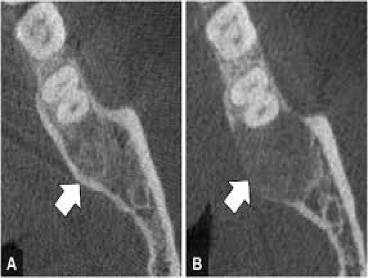
Conventional radiographs provide limited information due to their two-dimensional nature and superimposition of structures. CBCT overcomes these limitations by offering high-resolution, three-dimensional visualization of maxillofacial structures with relatively low radiation dose. CBCT enables detailed assessment of lesion extent, internal architecture, cortical involvement, and relationship with adjacent anatomical structures. In fibro-osseous lesions, CBCT typically reveals a spectrum of appearances ranging from radiolucent to mixed and radiopaque patterns, reflecting different stages of lesion maturation.
Image analysis of CBCT data allows quantitative evaluation of bone microarchitecture within fibro-osseous lesions. Parameters such as trabecular thickness, trabecular spacing, bone density distribution, and homogeneity can be assessed using specialized software. Fibrous dysplasia often demonstrates a characteristic “ground-glass” appearance with ill-defined borders and relatively uniform trabecular pattern, while ossifying fibroma tends to show a well-circumscribed lesion with mixed internal density and a more organized trabecular structure. Cemento-osseous dysplasia exhibits variable radiodensity depending on the stage, with immature lesions showing sparse trabeculation and mature lesions displaying dense mineralization.
CBCT-based image analysis also aids in evaluating cortical bone changes associated with fibro-osseous lesions. Expansion, thinning, or perforation of cortical plates can be accurately identified and measured. Assessment of cortical integrity is particularly important in determining lesion aggressiveness and planning surgical intervention. Additionally, changes in the surrounding cancellous bone microarchitecture may provide insight into lesion activity and progression.
Quantitative image analysis enhances diagnostic confidence by reducing subjectivity inherent in visual interpretation alone. By comparing microarchitectural parameters of the lesion with adjacent normal bone, clinicians can better differentiate fibro-osseous lesions from other radiographically similar entities such as chronic osteomyelitis, osteoma, or malignancies. Furthermore, longitudinal CBCT image analysis allows monitoring of lesion growth, maturation, or response to treatment, which is especially useful in conditions managed conservatively, such as fibrous dysplasia.
Despite its advantages, CBCT has limitations in absolute bone density measurement due to variability in gray values and lack of standardized Hounsfield units. However, relative comparisons within the same scan and standardized acquisition protocols can provide meaningful and reproducible data. Integration of CBCT with advanced image processing and artificial intelligence algorithms holds future promise for automated characterization and classification of fibro-osseous lesions.
In conclusion, CBCT-based evaluation of bone microarchitecture using image analysis offers a comprehensive, three-dimensional, and quantitative approach to the assessment of fibro-osseous lesions of the jaws. This technique enhances diagnostic accuracy, aids in treatment planning, and facilitates longitudinal monitoring, making it an invaluable tool in contemporary maxillofacial imaging.
No Any Replies to “CBCT-Based Evaluation of Bone Microarchitecture in Fibro Osseous Lesions Using Image Analysis”
Leave a Reply