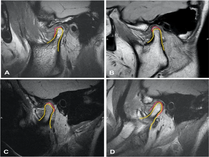
Magnetic Resonance Imaging (MRI) is a valuable tool in assessing the temporomandibular joint (TMJ) due to its ability to provide detailed images of soft tissues with excellent contrast resolution. MRI findings of the TMJ can reveal various pathologies and abnormalities, aiding in diagnosis and treatment planning for patients with temporomandibular joint disorders (TMD).
One of the most common indications for TMJ MRI is the evaluation of internal derangements, which encompass a range of conditions involving displacement or dysfunction of the articular disc within the joint. MRI can depict the position of the disc relative to the condyle during various phases of mandibular movement, helping to diagnose disc displacement with or without reduction. In disc displacement with reduction, the disc may be anteriorly displaced during mouth opening but reduces back onto the condyle upon closing. Conversely, in disc displacement without reduction, the disc remains anteriorly displaced, leading to symptoms such as pain, clicking, or limited mouth opening.
MRI can also identify degenerative changes within the TMJ, such as osteoarthritis or osteoarthrosis, characterized by joint space narrowing, subchondral sclerosis, osteophyte formation, and irregularity of the articular surfaces. These findings may correlate with clinical symptoms of joint pain, crepitus, and reduced range of motion.
Inflammatory conditions affecting the TMJ, such as synovitis or capsulitis, can be visualized on MRI as joint effusion, thickening of the synovial lining, and enhancement of soft tissues surrounding the joint. These findings may indicate active inflammation and can guide treatment decisions, such as the use of anti-inflammatory medications or intra-articular injections.
MRI is also sensitive to detecting abnormalities of the articular surfaces and surrounding structures, including bone marrow edema, subchondral cysts, and erosions. These findings may suggest traumatic injury, osteonecrosis, or inflammatory arthritis affecting the TMJ.
Furthermore, MRI can assess the integrity of the TMJ ligaments, including the collateral ligaments, lateral ligament, and sphenomandibular ligament. Ligamentous injuries, such as sprains or tears, may result from trauma or chronic joint hypermobility and can contribute to instability and dysfunction of the TMJ.
In addition to providing diagnostic information, MRI is useful for planning surgical interventions or other therapeutic procedures for TMJ disorders. For example, MRI can guide arthroscopic surgery by identifying the location and extent of disc displacement, as well as assessing the condition of the articular surfaces and surrounding soft tissues.
In summary, MRI findings of the TMJ encompass a wide range of pathologies and abnormalities, including internal derangements, degenerative changes, inflammatory conditions, articular surface abnormalities, and ligamentous injuries. By providing detailed anatomical information with excellent soft tissue contrast, MRI plays a crucial role in the diagnosis, treatment planning, and management of patients with temporomandibular joint disorders.


No Any Replies to “Magnetic Resonance Image Findings of TMJ”
Leave a Reply